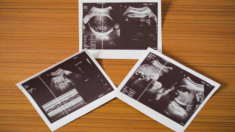
In the earlier era, there was less or no use of imaging during pregnancy and the babies were born without having any prior knowledge of any abnormality. But since the second half of the 20th century history of obstetrics has been revolutionized by the introduction of high-frequency transducers with real-time ultrasonography.
In this article, we will be discussing the different types of ultrasound, how to read and interpret ultrasound, and the colors which are seen there.
What Is An Ultrasound Scan?
Ultrasonography is a procedure that uses high frequency ( 2 to 10 MHz) of sound waves which in turn creates images on the screen by converting the reflective images to the visual images. Now, these sound waves are being emitted through the transducers which are used during the scan. The real-time images that are seen on the screen, it is produced by the sound waves which in turn reflect from fluid and tissues of the fetus.
What Are The Different Types of Ultrasound In Pregnancy?

1. Transvaginal ultrasound
Transvaginal ultrasound in pregnancy is done only in the first trimester or during the early gestation till 10 weeks. Here, the probe is introduced into the patient’s vagina for getting a clear image. The bladder should be empty and the frequency of the probe is between 5 to 9 MHz. Since in the earlier weeks via transabdominal scan it is difficult to visualize the fetus properly, so transvaginal ultrasound is preferred.
2. Standard ultrasound
Standard ultrasound generally means which is done via the transabdominal probe. It is generally done in the second and third trimester of pregnancy. Here the probe has a frequency of 2 to 5MHz and it needs to be performed with a full bladder for good visualization.
The standard scans are generally done by booking around 11 to 14 weeks of gestation to identify any genetic abnormality which is called a nuchal translucency scan. The next booking scan is to be done between 16 to 20 weeks of pregnancy which determines any abnormality in the structural development of the fetus.
3. Fetal echocardiography
Fetal echocardiography generally is done between the 23 to 25 weeks period of gestation. It is a detailed study of the fetal cardiac anatomy and the flow pattern. This is generally done via a 2D probe, but in recent advancements, it is replaced by 3D/4D transducers. It is done by analyzing the color flow and spectral analysis through all four chambers of the heart by Doppler.
The scan is generally done by a specialized and trained person like a pediatric cardiologist. It is not routinely done since it is costly, only done if the patient has a previous history of cardiac anomalies in a previous pregnancy, familial history of genetic cardiac disease, or even earlier scan showing any suspected cardiac anomalies. (1)
4. 3-D ultrasound
3D ultrasound has been made popular In the last two decades, but it is not routinely used during the standard ultrasound of pregnancy. It is a component of various special evaluations. The 3D scan is done by a specialized transducer. It is used for specialized images in different planes of the fetus. Though not commonly used but can detect any abnormality or anomaly of the fetus in utero. Sometimes, parents wish to visualize the face of the fetus which cannot be visualized properly by 2D ultrasound. It is also used to detect any condition of the fetus which is around the amniotic fluid. (2)
5. Dynamic 3-D ultrasound (4D ultrasound)
It is also called the real-time imaging of the fetus in utero. This ultrasound is the repetition of the 3D images multiple times. It is generally used to detect specific clinical abnormalities like cleft lip, cleft palate, brain anomalies, etc. This advanced form of ultrasound is not routinely done in India, generally in Western countries it is being used regularly (3).
How to Read an Pregnancy Ultrasound Report?

1. Look at your womb
While performing the ultrasound or for the new parents when the doctor gives the picture of the ultrasound location of the womb, which means where the baby is present needs to be identified. The white or gray lines in the scan are the uterus inside which the black-colored sac-like structure is seen, that is the amniotic fluid where the small baby is present. But again the image can change the position of the baby according to the placement of the probe by the doctor.
2. Look at your baby
After identifying the womb, the next important structure to notice is the baby, which can be seen as a white or grayish image. The size of the baby can vary according to the gestation of the pregnancy. They are:
- Early gestation till around 8 to 10 weeks, the baby or the fetus can be seen as a lemon seed or bean.
- Following 12 weeks to 20 weeks of gestation the baby can be seen with a small head, spine, hands, and leg.
- Following 20 weeks till delivery, the baby can be visualized properly with proper face, eyes, nose, spine, hands, and legs.
How To Interpret Different Colors In A Pregnancy Ultrasound Report?
Traditional images are black and white but colors can also be seen there which can be seen via the color Doppler option. The color coding seen in ultrasound is to assess the direction of blood flow and the speed of the flow between the blood vessels. (4)
Typically seen colors in ultrasound are:
- Red: it indicates that the blood flow is towards the probe.
- Blue: it indicates the blood flow is away from the probe
- Green: this generally indicates the velocity of the blood flow is low
- Yellow or orange: it indicates the turbulence that is created in the blood flow. (5)
The ultrasonography procedure and the interpretation is not that easy to understand. It needs expertise to properly interpret the ultrasound sound report. So parents need to ask and consult with the doctor for proper diagnosis of any problem detected.
What Are Normal Pregnancy Ultrasound Results?

The ultrasound results in pregnancy vary according to the gestational age and the trimester we are looking into .(6)
First trimester
A scan done in this period is to look for the gestational age, location of pregnancy, single/ twin/ triplet, and calculate the estimated delivery date.
Second trimester
In this period the organ development, any anomalies, cardiac ultrasound, and gender of the fetus are determined.
Third trimester
This is the last and the crucial monitoring phase of pregnancy. Here amniotic fluid volume, Doppler for proper blood flow, estimated fetal weight, and growth are determined.
FAQ’s
1. How Can You Tell On An Ultrasound If It Is A Boy Or a Girl?
The fetal gender can be determined between 12 to 16 weeks of gestation. Though it is illegal in India and it is a punishable offense, in Western countries it is regularly practiced. By doing 3D and 4D ultrasound it can be identified that the fetus is a boy or a girl if both the legs of the fetus are widely apart.
References
- Rychik J, Ayres N, Cuneo B, Gotteiner N, Hornberger L, Spevak PJ, Van Der Veld M. American Society of Echocardiography guidelines and standards for performance of the fetal echocardiogram. Journal of the American Society of Echocardiography. 2004 Jul 1;17(7):803-10 – https://pubmed.ncbi.nlm.nih.gov/15220910/
- MERZ, EBERHARD MD*; ABRAMOWICZ, JACQUES S. MD†. 3D/4D Ultrasound in Prenatal Diagnosis: Is it Time for Routine Use?. Clinical Obstetrics and Gynecology 55(1):p 336-351, March 2012. | DOI: 10.1097/GRF.0b013e3182446ef7 – https://pubmed.ncbi.nlm.nih.gov/22343249/
- Pooh R, Maeda K, Kurjak A, Sen C, Ebrashy A, Adra A, Dayyabu A, Wataganara T, de Sá R, Stanojevic M. 3D/4D sonography – any safety problem. Journal of Perinatal Medicine. 2016;44(2): 125-129 – https://www.degruyter.com/document/doi/10.1515/jpm-2015-0225/html
- Malik R, Saxena A. Role of colour Doppler indices in the diagnosis of intrauterine growth retardation in high-risk pregnancies. The Journal of Obstetrics and Gynecology of India. 2013 Feb;63:37-44 – https://www.ncbi.nlm.nih.gov/pmc/articles/PMC3650155/
- Alfirevic, Z., & Kurjak, A. (1990). Transvaginal colour Doppler ultrasound in normal and abnormal early pregnancy. Journal of perinatal medicine, 18(3), 173–180. https://doi.org/10.1515/jpme.1990.18.3.173 – https://pubmed.ncbi.nlm.nih.gov/2200863/
- Hsu, S., & Euerle, B. D. (2012). Ultrasound in pregnancy. Emergency medicine clinics of North America, 30(4), 849–867 – https://pubmed.ncbi.nlm.nih.gov/23137399/
