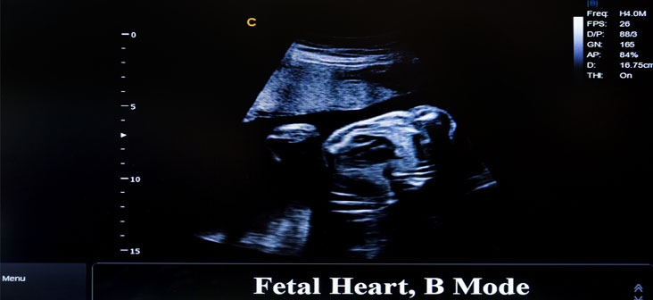
Some pregnant women are at a higher risk of giving birth to a baby with congenital heart disease. About 1% of babies are born with heart issues ranging from mild to severe. It is highly beneficial if the CHD is diagnosed when the baby is in the womb itself. The prenatal diagnosis of CHD is found to improve the outcome of pregnancy. This is because prenatal diagnosis allows the baby to have faster access to medical and surgical intervention (if needed) soon after birth and also help the pregnant woman to better manage the pregnancy and delivery in case of any abnormality. Fetal echocardiography is one such procedure with which CHD of the fetus is diagnosed before the delivery. Read on to understand more about this method.

What Is Fetal Echocardiography?
Fetal echocardiography is a particular ultrasound scan used for the detailed study of fetus’s heart for issues and abnormalities like valves that do not open and close properly, hole in the heart, narrowing arteries, the heart rhythm disturbances etc. Fetal echocardiography evaluates the heart’s shape, structure and function.
When Is Fetal Echocardiography Recommended?
Fetal echocardiography is not a routine procedure in all pregnancies. For most pregnancies, a basic ultrasound, which is the part of prenatal care, will show whether all four chambers of the baby’s heart have developed. The obstetrician will recommend conducting this procedure under the following circumstances:
- If the mother has some health issues like type 1 diabetes (insulin dependent), phenylketonuria –an inability to break down an essential amino acid called phenylalanine, lupus, connective tissue disease etc. which puts the pregnancy in a high-risk category
- If a routine prenatal scan reveals some abnormalities with the structure or function of the heart
- If abnormal fetal heart rhythm is noticed
- If an amniocentesis detects some chromosomal or genetic disorder
- If the mother to be consumed alcohol or used drugs during pregnancy
- If there is a family history of congenital heart disease
- If the pregnant woman has congenital heart disease
- If the mother to be has already given birth to a baby with congenital heart disease
- If the pregnant woman was on any medication that may affect the normal development of the fetal heart and increase the risk of the fetus to have congenital heart disease. Medicines for epilepsy, severe acne (like Accutane), etc. are some examples
- If the fetus possesses twin-to-twin transfusion syndrome
- If increased nuchal translucency (3.5mm or more) is detected on the first-trimester screening
When Is Fetal Echocardiogram Performed?
Fetal echocardiogram is performed during the second trimester, precisely between 18 to 24th week.
Preparation For The Procedure
There is no preparation required for taking the test. You will also not be required to take the test on a full bladder as in case of prenatal ultrasound.
The duration of the test will vary from 30 minutes to 2 hours depending on the situation. Therefore it is recommended to have someone like your spouse or a family member accompany you for the test

How Is A Fetal Echocardiogram Performed?
A specially trained ultrasound sonographer usually performs the fetal echocardiogram and a pediatric cardiologist who specializes in fetal congenital heart disease interprets the images that are obtained. The procedure is similar to a prenatal ultrasound. The pregnant woman is required to lie down for the procedure. There are two ways to perform a fetal echocardiogram:
- Abdominal ultrasound: This is the most widely recognized type of ultrasound to assess the fetus’s heart. A gel like solution is spread on the pregnant woman’s belly. A handheld ultrasound probe is gently placed on her belly and will be moved around to obtain images of various area, structure and shape of the fetal heart on a computer screen. This test causes no pain or harm to the mother to be or her unborn baby. The test takes normally 30–120 minutes relying upon the complexity of the baby’s heart
- Endo vaginal or transvaginal ultrasound: A transvaginal echocardiography is normally done during the early stages of pregnancy. A much smaller probe is placed into the vagina and leans against the back of the vagina. The pictures of the fetus heart are captured. The picture delivered by transvaginal echocardiography will be clearer than an abdominal ultrasound
Some other techniques that are used to get more clear and detailed information about the fetal heart includes:
- 2D Echocardiography: This technique is used to evaluate the actual size, structures and movement of the fetal heart
- Doppler echocardiography: It is a method used to assess the blood flow between the heart valves and chambers. This technique can easily detect abnormal blood flow inside the heart, which points out problems like an issue with any of the valves, or a problem in the fetal heart wall etc
- Color Doppler: This is an improved version of doppler echocardiography. With color doppler, different colors are used to indicate the blood flow direction. This makes interpreting the doppler images much simpler
What Do The Test Results Indicate?
During your follow up appointment, your doctor will interpret and communicate the test results to you. The test results will have the following meaning:
- If there is no cardiac abnormality found, then the test results will be normal
- If there is some abnormality found like abnormal rhythm, heart defect or any other problem in the test results, then the doctor may ask the pregnant woman to undergo additional tests like a fetal MRI or other ultrasounds to detect the exact issue
Risk Associated With Fetal Echocardiography
There are no risk associated with the echocardiogram since it utilizes ultrasound rather than radiation.
What Are The Limitations Of Fetal Echocardiography?
Fetal echocardiography cannot be used to diagnose every possible abnormality. If there is a hole in the heart or mild issues with valves, fetal echocardiogram will not be able to detect it. Sometimes even the abnormal results can be inconclusive so if your doctor feels the necessity, he may ask you to repeat the test or undergo additional tests.
Read Also: Electronic Fetal Monitoring During Labor and Delivery

