 Anencephaly is a rare but devastating neural tube defect that occurs during pregnancy, where a major portion of a baby’s brain and skull fail to develop. Among neural tube defects, anencephaly had the highest prevalence at 2.1 per 1000 births followed closely by spina bifida at 1.9 per 1000 births. (1). Thus, it is important to understand what causes anencephaly during pregnancy.
While the exact cause remains unknown, research points to a combination of genetic, environmental, and lifestyle factors. Early prevention, through proper prenatal care, folic acid supplementation, and avoiding certain risks, plays a crucial role in reducing its occurrence. Let’s dive into the key contributors and explore the science behind Anencephaly.
What is Anencephaly?
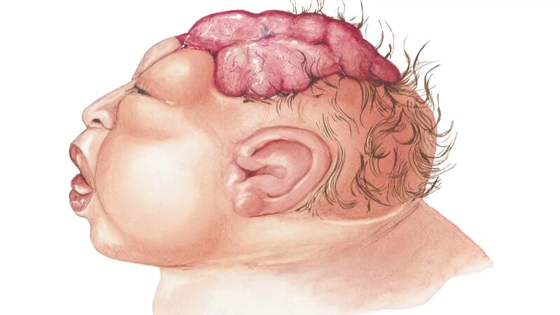
Anencephaly is a severe birth defect in which a baby is born without parts of the brain and skull. It occurs when the upper portion of the neural tube, which eventually forms the brain and spinal cord, fails to develop properly during early pregnancy, typically within the first four weeks. As a result, the baby is often born without a significant portion of the brain, including the cerebrum. The cerebrum controls thinking, motor skills, and senses. Most babies with anencephaly are either stillborn or die shortly after birth. (2)
Anencephaly is part of a group of defects known as neural tube defects (NTDs). While the exact cause is still not fully understood, it is believed that a combination of genetic and environmental factors contributes to its development. Pregnant women who don’t get enough folic acid—an essential vitamin for proper fetal development—are at a higher risk of having a baby with this condition.
What Are The Different Types Of Anencephaly?
Typically, there are three types of anencephaly, such as:
- Meroanencephaly: The midbrain and brainstem are partially developed and skin cover and skull partially cover the brain.
- Holoanencephaly: In this condition, the brain of the baby is completely absent. Holoprosencephaly is a common form of anencephaly.
- Craniorachischisis: In this type of anencephaly, the brain, skull, and spine of the newborn baby are absent. Craniorachischisis is a severe anencephaly type and generally, it is a combination of a spine defect and anencephaly.
What Are The Signs Of Anencephaly?
Some of the common signs of anencephaly are as follows:
- Folded Ears: The baby will have folded ears.
- Skull Bones Absent: Bony covering may be absent from the back of the head. In some cases, bones are also missing from the side and front of the head.
- Cleft Palate: The roof of the baby’s mouth may not close properly, leaving an opening that may also extend to the nasal cavity.
- Flattened Head: Babies’ heads may seem flattened due to incomplete brain development and absent skull bones.
- Congenital Heart Defects: Babies born with this condition may have congenital heart defects or structural problems with their valves, chambers, and walls.
- Polyhydramnios: In this condition, the amniotic sac might be filled with excessive amniotic fluid.
- Smaller Head Size: Babies with anencephaly conditions may have a smaller head than regular size.
- Brain Tissue Exposed: The tissue of the brain may be exposed due to the absence of bones and skin.
What Causes Anencephaly in Babies?
Although there are no well-researched and defined reasons behind anencephalic babies, a few factors falling under the category of genetics, environment, and nutrition during pregnancy have been identified and discussed below:
- Anencephaly or exposed brain syndrome, is not hereditary. But if the previous pregnancy of the lady did have an anencephalic fetus, there is a high chance of having the next pregnancy with anencephaly too.
- Lack of folic acid in women during pregnancy can lead to anencephalic baby development within her. So pregnant ladies are often advised to take prenatal vitamins containing 400 micrograms of folic acid during and before pregnancy. Those who don’t get enough folic acid (vitamin B9) when they’re pregnant have a higher risk of having a baby with anencephaly.
- The impact of gestational diabetes on the baby is well known. Uncontrolled gestational diabetes increases the risk of neural tube defects. The high content of glucose in the blood does affect fetal development. (3)
- Making use of a sauna bath or hot water tub during the early stages of pregnancy puts the pregnant lady at risk of giving birth to anencephalic babies.
- An excessive dosage of anti-seizure drugs like phenytoin, carbamazepine, and valproic acid can cause NTDs. (4)
- Obesity and pregnancy complications go hand in hand. Obese pregnant ladies are at higher risk of having an anencephalic baby.
- Consumption of opioids during the initial two months of pregnancy can lead to NTDs. Opioids consist of heroin, which is an illegal drug, and hydrocodone.
Read: Is It Safe To Take Hydrocodone During Pregnancy?
What Are The Symptoms Of Anencephaly?
Although the kind of symptoms of an anencephalic baby vary from individual to individual, a few diagnosed symptoms are mentioned here below:
- Lack of bone on the back of the head
- Absence of bones in the front and sides of the head
- Absence of cerebrum in brain
- Folded ears
- Split in the cleft palate of the mouth
- Congenital heart defects
Risk Factors for Anencephaly
Some risk factors for anencephaly include:
- Deficiency of folic acid (vitamin B9) before pregnancy and during pregnancy. (5)
- Prolonged medication for such conditions as migraine headaches, seizures, and bipolar disorder.
- High exposure to heat during pregnancy, such as using a sauna or hot tub and having a consistent fever.
- Opioid usage during pregnancy.
- Diabetes during pregnancy.
Diagnosis and Treatment For Anencephaly
Anencephaly is a condition that cannot be treated, which makes it a fatal form of neural tube defect (NTD). During pregnancy, the healthcare providers might recommend the pregnant lady a few tests to look for symptoms related to neural tube defects.
Prenatal tests for anencephaly include:
1. Quad Marker Screen Blood Test
In this blood test, the neural tube defects and genetic disorders present in the fetus can be detected. If in the test report, the level of alpha-fetoprotein (AFP) is high, there are chances of getting an anencephalic fetus.
2. Ultrasound
During a prenatal ultrasound, the pictures of the unborn baby look at the baby’s skull, brain, and spine. In case of non-formation of part of the brain, the anencephalic fetus can easily be identified.
3. Fetal Magnetic Resonance Imaging (MRI)
In case of doubt regarding the presence of anencephalic syndrome, the healthcare provider may ask for an MRI scan to get a clear picture of the brain and spine in greater detail.
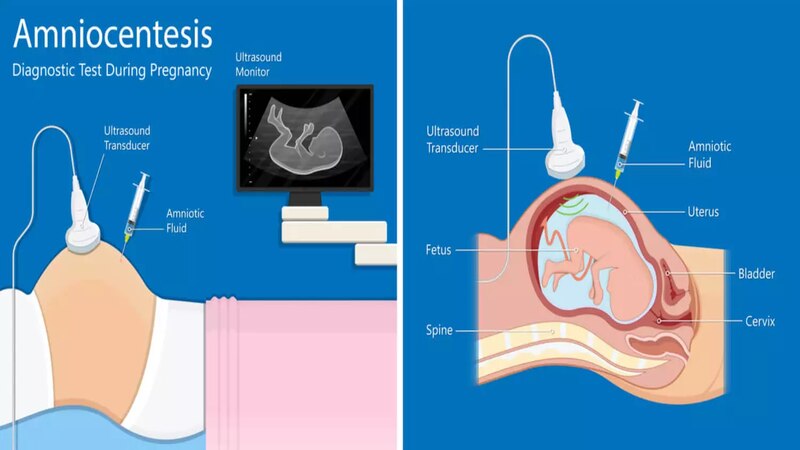
4. Amniocentesis
In this test, a fine needle is inserted into the amniotic sac to take a sample of the amniotic fluid. Thereafter, the level of AFP is tested in amniotic fluid. In the case of high levels of AFP or acetylcholinesterase enzyme, it is confirmed that the fetus is anencephalic.
Can Anencephaly Be Inherited?
No, typically anencephaly is not inherited (not passed from family traits). It can occur without any family history of genetic conditions, such as sporadic genetic mutation. However, suppose you previously had a baby born with a neural tube defect such as spina bifida. In that case, you are likely to have higher chances of giving birth to a baby with anencephaly.
What Are The Features Of Anencephaly After Ultrasound?
Anencephaly is a condition when a baby is born without a significant portion of the brain, skull, and skin. After ultrasound, the features of anencephaly are as follows:
- Top of the Skull is Absent: In ultrasound, the bones that form the top of the skull appear to be missing.
- Underdeveloped or Missing Brain Part: In ultrasound, the brain seems either underdeveloped or some parts are missing.
- Frog Eye: In some cases, the ultrasound may show the characteristic appearance that resembles a frog eye.
- Polyhydramnios: The presence of excessive amniotic fluid may be seen in the ultrasound report.
Is Anencephaly in Babies Preventive?
The neural tube (NT) closes twenty-eight to thirty-two days after a baby is conceived. Many women are not even aware of their pregnancy during these initial days. It is during the initial three to eight weeks of pregnancy that the neural tube gets affected by factors such as
- Lack of folic acid and other nutrients
- Infection
- Usage of medicine or alcohol
- A hazardous chemical-infused environment surrounding
- Any genetic disorder
Anencephaly is a devastating condition where many affected babies are stillborn or die shortly after birth, often within hours or days. In rare cases, if a baby survives for weeks, it may lose the ability to respond to sound or touch, becoming unconscious, deaf, or blind. Women who carefully maintain optimal folic acid levels during pregnancy significantly reduce the risk of anencephaly. To ensure this, pregnant women should focus on eating leafy greens, citrus fruits, nuts, beans, and fortified cereals to boost their folic acid intake. Even if you’re not pregnant but in your childbearing years or planning to conceive, start taking folic acid and vitamins to minimize the risk of anencephaly. If a healthcare provider detects a neural tube defect, an increased folic acid dosage is recommended before and after conception.
FAQS
1. What Is The Cause Of Anencephaly?
Anencephaly is a form of birth defect that typically occurs when the neural tube remains half opened or doesn’t close completely during pregnancy.
2. Can Babies Survive With Anencephaly?
No, generally babies with anencephaly do not survive. It is a severe birth defect that causes a baby to be born without a significant part of the brain, scalp, and skull. Moreover, babies with this condition are stillborn and usually live for a few hours only.
3. Is Anencephaly Curable?
No, anencephaly is a non-curable condition. It is a fatal birth defect and babies with anencephaly are born without brain and skull parts.
4. What is The Longest Living Baby With Anencephaly?
The longest-living baby with anencephaly survives till 28 months without any life-sustaining treatment.
References
- Bhide P, Sagoo GS, Moorthie S, Burton H, Kar A. Systematic review of birth prevalence of neural tube defects in India. Birth Defects Res A Clin Mol Teratol. 2013 Jul;97(7):437-43 – https://pubmed.ncbi.nlm.nih.gov/23873811/
- Madrid L, Vyas KJ, Kancherla V, Leulseged H, Suchdev PS, Bassat Q, Sow SO, El Arifeen S, Madhi SA, Onyango D, Ogbuanu I, Scott JAG, Blau D, Mandomando I, Keita AM, Gurley ES, Mahtab S, Akelo V, Sannoh S, Tilahun Y, Varo R, Onwuchekwa U, Rahman A, Adam Y, Omore R, Lako S, Xerinda E, Islam KM, Wise A, Tippet-Barr BA, Kaluma E, Ajanovic S, Kotloff KL, Hossain MZ, Mutevedzi P, Tapia MD, Rogena E, Moses F, Whitney CG, Assefa N; CHAMPS Consortium – https://pmc.ncbi.nlm.nih.gov/articles/PMC10282076
- Sean M. Tafuri; Forshing Lui. California Northstate University; 2 CA Northstate Uni, College of Med – https://www.ncbi.nlm.nih.gov/books/NBK545244/
- Nie Q, Su B, Wei J. Neurological teratogenic effects of antiepileptic drugs during pregnancy. Exp Ther Med. 2016 Oct;12(4):2400-2404 – https://pmc.ncbi.nlm.nih.gov/articles/PMC5038337/
- CDS, Neural Tube Defects – https://www.cdc.gov/birth-defects/about/neural-tube-defects.html